文章信息
- 陶丽, 朱才丰, 葛宏慧, 等.
- TAO Li, ZHU Caifeng, GE Honghui, et al.
- 艾灸督脉对阿尔茨海默病患者认知功能及血浆外泌体中Aβ1-42和lncRNA H19的影响
- Effects of moxibustion on Du meridian in cognitive function and plasma exosomes Aβ1-42 and lncRNA H19 in Alzheimer's patients
- 天津中医药, 2023, 40(7): 864-870
- Tianjin Journal of Traditional Chinese Medicine, 2023, 40(7): 864-870
- http://dx.doi.org/10.11656/j.issn.1672-1519.2023.07.09
-
文章历史
- 收稿日期: 2023-04-03
2. 安徽中医药大学第二附属医院老年病三科,合肥 230061;
3. 上海市闵行区颛桥社区卫生服务中心,上海 201100
阿尔茨海默病(AD)在《景岳全书》中属“痴呆”范畴,该病是以记忆力减退为特征的慢性神经退行性疾病,好发于老年群体。目前该病发病机制主要有淀粉样蛋白级联假说、tau蛋白过度磷酸化假说等。研究表明Aβ1-42易聚集在海马、大脑皮质中,与AD相关性更强[1]。本课题组前期研究发现,通过下调APP/PS1双转基因小鼠海马组织长链非编码RNA H19(lncRNA H19)表达,可改善AD小鼠自噬-溶酶体功能,促进细胞自噬清除脑内Aβ,改善认知功能[2]。当前有大量临床研究表明中医治疗AD有效[3-4],但对治疗的客观指标变化观察较少。
本次研究观察艾灸督脉组穴治疗AD患者的临床疗效,同时探索治疗前后血浆外泌体中Aβ1-42浓度和lncRNA H19的表达变化,为艾灸督脉改善AD患者认知功能提供客观依据。
1 资料与方法 1.1 病例来源及分组方法研究对象为2021年1—12月安徽中医药大学第二附属医院老年病三科门诊、病房收治的30例阿尔茨海默病患者为AD组。同时招募30例年龄、性别匹配,且无痴呆病史的健康人作为健康对照组。本研究遵照《赫尔辛基宣言》,通过了安徽中医药大学第二附属医院伦理委员会审查(编号:2021zj38)。
1.2 诊断标准参照2011年美国国家老龄化研究院和阿尔茨海默病协会发布的诊断标准(NIA-AA诊断标准)[5]。采用临床痴呆评定量表(CDR)评定患者痴呆程度,得分为0、0.5、1、2和3分,分别代表健康、可疑、轻度、中度和重度[6]。(CDR是一个用于痴呆诊断和严重程度分级的临床总体评价量表,在全球范围内广泛用于流行病学研究、病例分析和临床试验[7]。)
1.3 纳入标准1) 年龄50~80岁,性别不限,病程在6个月至3年。2)符合上述诊断标准。3)CDR评分1~3分。4)所有患者既往无精神病史、癫痫史、无其他器官疾病史。5)自愿参与本次研究并签署知情同意书。
以上为AD组纳入标准,健康对照者须无痴呆病史,年龄50~80岁,且不符合AD诊断标准。
1.4 排除标准1) 排除鉴别诊断中的的其他痴呆,如血管性痴呆、额颞叶痴呆等。2)患有其他严重疾病者。3)无法配合研究者。4)存在药物过敏史、出血性疾病者。
1.5 脱落标准1) 不符合纳入标准,被误纳入者。2)纳入后不能配合治疗或中途停止治疗的患者。3)擅自进行其他治疗方案,对本次试验有效性和安全性产生影响的患者。
1.6 艾灸方法参照人民卫生出版社第3版《针灸学》所述方法取“百会”“大椎”“风府”“命门”。AD患者取坐位,医者将直径2.5 cm,厚度0.5 cm的附子饼用针穿刺数个小孔后放置在百会穴上,点燃清艾条,一手拇指和食指呈“C”形固定附子饼,另一手将艾条燃烧端实按于附子饼上,待患者诉皮肤灼痛时立即抬起艾条片刻,如此反复施灸。将艾条燃烧端悬于大椎、风府、命门穴上方施以温和灸,以患者自觉皮肤灼热、可耐受为度。每穴各灸20 min,每日1次,治疗8周。
1.7 不良事件的观察、处理及记录对试验期间出现的不良事件,详细记录并评估与试验的相关性。在试验过程中若发生严重不良事件,应立即中止试验并对受试者采取适当的治疗措施,详细记录停止时间、不良事件发生时间、处理经过及转归,并上报。
1.8 疗效指标与疗效评价采用MoCA量表和ADAS-cog量表评估患者认知功能。MoCA量表满分30分,评分≥26分为认知功能正常,若测试者受教育的年限不超过12年,则总分加1分[8]。ADAS-cog量表范围在0~75分,分数越高认知功能越差[9]。两者均为AD评价工具,前者具有高度的区分度和筛查价值,后者在评估AD人群方面具有良好条目相关性和结构效度[10]。
采用《老年呆病(老年痴呆)的中医临床诊断及疗效评定标准(试行)》的相关标准[6]。
观察疗效:1)基本痊愈:积分﹥64分。2)显效:积分增加﹥16分。3)有效:积分增加﹥9分。4)无效:积分增加<8分。5)恶化病情加重:积分减少或死亡。
1.9 血浆外泌体Aβ1-42和LncRNAH19的测定收集健康受试组和治疗前后AD组的血液样品各5 mL,放入离心机中(离心半径:13 cm;离心速度:3 000 r/min;离心时间:5 min)离心。再将获得的血浆样本过滤、重悬,得到外泌体。取10 μL外泌体滴在铜网上,沉淀1 min后,用滤纸吸去浮液,在干净铜网上滴10 μL醋酸双氧铀,沉淀1 min后,再次滤纸吸去浮液,常温干燥后电镜检测成像,利用NTA软件结合NS500纳米颗粒分析仪,对外泌体溶液进行纳米粒子追踪分析和外泌体定量检测。再使用酶联免疫吸附试验(ELISA)试剂盒对外泌体样本的特异性标志蛋白CD81进行检测[11],鉴定外泌体及对Aβ1-42进行测定。最后通过外泌体RNA的提取、DNA消除、反转录获得cDNA,再以cDNA为模板通过Real-time荧光定量聚合酶链反应(PCR)扩增,最后以相对表达量2-ΔΔCt计算目的基因的相对表达量。
比较AD组艾灸干预前后与健康受试组相比血浆外泌体中Aβ1-42蛋白浓度与lncRNA H19表达量变化。
1.10 统计学方法采用SPSS 26.0统计学软件进行统计分析,计量资料采用均数±标准差(x±s)表示。组内治疗前后比较,差值d符合正态分布者采用配对t检验,不符合正态分布则采用配对符号秩和检验;两组间采用独立样本t检验。相关性采用Pearson线性相关分析。P<0.05为差异有显著性意义。
2 结果 2.1 一般情况本次研究共60例符合标准的人员参与试验,AD组在治疗期间无不良事件发生、无病例脱落。最终两组各30例资料纳入分析。两组数据一般资料均无统计学差异,具有可比性(P>0.05)。见表 1。
AD组治疗后MoCA评分较治疗前有提高(P < 0.05),ADAS-cog量表评分较治疗前有降低(P < 0.05)。见表 2。AD患者治疗前后各等级的分布情况见表 3,AD患者疗效比较见表 4。
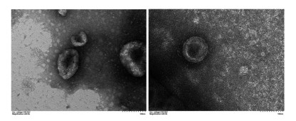
在透射电镜下观察到提取的物质呈杯托盘状或囊泡状,膜性结构,大小均匀、形态一致,直径约40~100 nm,可聚集成群,也可单个分布。见图 1。
|
| 图 1 外泌体在透射电镜(100 nm)下的形态 Fig. 1 Morphology of exosomes under transmission electron microscopy (100 nm) |
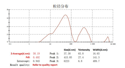
NS500仪器下观察见呈布朗运动的悬浮纳米颗粒,对纳米颗粒进行粒径分析,绘制颗粒分布曲线,结果显示颗粒平均粒径为58.19 nm,强度为68.8%。见图 2。
|
| 图 2 外泌体粒径分析图 Fig. 2 Exosome particle size analysis |
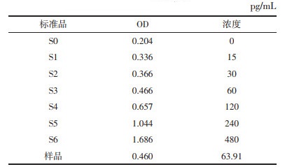
用ELISA的方法对不同浓度的标准品蛋白浓度在OD 450 nm处的吸光度值的结果,得到标准曲线方程:Y=0.003X+0.273 7,r2=0.994 4。该实验验证了外泌体的测定。见表 5。
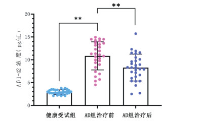
AD组治疗前与健康对照组、治疗前与治疗后血浆外泌体Aβ1-42浓度比较,差异均有统计学意义(P < 0.01)。见图 3。
|
| 注:AD为阿尔茨海默病。每一个点代表每一个值,蓝色代表健康对照组,红色代表AD组干预前,紫色代表AD组干预后。**P < 0.01。 图 3 血浆外泌体Aβ1-42浓度水平比较 Fig. 3 Comparison of plasma exosome Aβ1-42 concentrations |
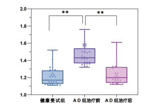
AD组治疗前与健康对照组、治疗前与治疗后血浆外泌体lncRNA H19比较,差异有统计学意义(P < 0.01)。见图 4。
|
| 注:每一个点代表每一个值,蓝色代表健康对照组,紫色代表AD组干预前,红色代表AD组干预后。**P < 0.01。 图 4 血浆外泌体lncRNA H19相对表达量水平比较 Fig. 4 Comparison of relative expression levels of plasma exosome lncRNA H19 |
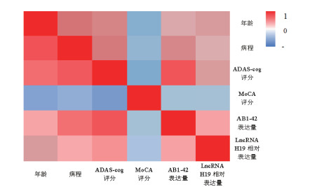
利用Pearson相关性分析得到对应的相关系数,显示年龄、病程与血浆外泌体Aβ1-42浓度和lncRNA H19相对表达量存在正相关性(P<0.05);ADAS-cog评分与Aβ1-42浓度水平、lncRNA H19相对表达量存在正相关性(P < 0.05);MoCA评分与Aβ1-42浓度水平、lncRNA H19相对表达量存在负相关性(P < 0.05);MoCA评分与ADAS-cog评分存在较强的负相关性(P < 0.05)。见图 5。
|
| 注:红、蓝分别代表正、负相关,颜色越深,相关系数绝对值越大。 图 5 AD组各指标间的相关性 Fig. 5 Correlation between indicators in AD group |
目前中国老龄人口与日俱增,AD发病率随之增加[12]。研究表明,2025年受该病影响的人数在全球范围内将达到1.2亿[13]。在中医基础理论中,髓海渐空、肾阳亏虚为AD发病之本。本次研究艾灸百会穴,以期醒脑开窍、通督调神之效,协同大椎、风府、命门,共奏补肾填髓之功。AD的西医治疗作用机制单一、疗效局限;中医治疗则强调多靶点、科学整合[14-15]。生理上,督脉行经人体后正中线及头部,与肾、脑直接相联系。本团队蔡圣朝主任从“神-脑-督脉-肾-任脉”轴入手,创立“温阳补肾灸”,有效预防及治疗痴呆[16]。在本团队前期研究中,以“温阳补肾灸”治疗轻度认知障碍(MCI)患者取得良好疗效,表明艾灸督脉可有效改善MCI患者的认知能力[17]。前期动物实验中也证实,艾灸督脉能上调细胞自噬水平、加速Aβ1-42清除、抑制炎性反应、促进坏死神经元修复再生,改善实验小鼠认知和记忆能力[18-21]。
MoCA量表是对认知功能异常进行快速筛查的评定工具,其敏感性高、覆盖重要认知领域、测试时间短,适合临床运用。包括注意与集中、执行功能、记忆等11个版块,总分30分,≥26分为正常认知水平[22]。本研究结果显示MoCA量表治疗后均值为20.83分,较治疗前18.76分有提高(P < 0.05)。ADAS-cog为痴呆干预研究实验的量表之一,是美国FDA推荐的临床试验认知评价的金标准[9]。包括语词回忆、命名、执行指令等12个条目,评分范围0~75分,评分越高表明认知受损越严重。本次研究显示AD组治疗后ADAS-cog量表均值为24.17分,较治疗前34.37分有明显降低(P < 0.05),提示认知功能在灸疗后得到改善。
长链非编码RNA (LncRNA) 是近来分子生物学的研究热点。长度超过200个核苷酸的lncRNA可通过表观遗传修饰与转录后加工等多种调控机制参与神经系统疾病的病理过程[23]。外泌体携带的遗传片段和毒性蛋白质可在细胞间运输,它参与神经元调节、细胞废物清除、免疫应答调节等,能被多数类型细胞释放[24-25],其内容物可发挥生物学功能[26]。CD81在外泌体中高度富集,是外泌体的常用标记蛋白[27-28]。研究表明,外泌体中携带的LncRNA H19可参与神经系统调节[29],抑制lncRNA H19表达和上调的miR-129可加速Aβ25-35刺激的PC12细胞的活力并抑制其凋亡,有利于AD的治疗[30]。研究表明,血浆中Aβ1-42水平能提前10年预测AD的临床发作[31]。在前期基础研究中也已证实,艾灸督脉能通过调控细胞自噬水平、促进Aβ清除等途径改善模型认知障碍大鼠的学习、记忆等认知能力[19-20, 32]。但脑脊液中Aβ蛋白的检测操作有创伤,导致临床使用受限,外泌体在体液中分布广泛,易于获得[33]。故从可行性角度分析,血浆外泌体检测存在优势。
本次研究发现,与健康受试组相比,AD组治疗前血浆外泌体Aβ1-42及lncRNA H19表达水平均较高;艾灸干预后,AD患者血浆外泌体中Aβ1-42浓度及lncRNA H19表达水平均有所降低。提示AD患者血浆外泌体Aβ1-42及lncRNA H19表达水平均较健康人高,艾灸干预对抑制血浆外泌体Aβ1-42及lncRNA H19表达水平作用确切。将血浆外泌体Aβ1-42、lncRNA H19的表达水平及临床观察指标进行相关性研究,发现血浆外泌体中Aβ1-42、lncRNA H19水平与ADAS-cog评分、MoCA评分具有一定相关性,提示提示血浆外泌体lncRNA H19及Aβ1-42可能作为AD患者生物学诊断和疗效评价的指标。
综上,艾灸督脉组穴能降低AD患者血浆外泌体中lncRNA H19的表达水平,促进Aβ1-42清除,改善患者认知能力。由于AD疾病中晚期治疗效果不理想,使得探索出早期诊断的特异性指标成为临床亟待解决的问题。而血浆外泌体Aβ1-42、lncRNA H19表达水平与AD相关性强,有望成为临床早期诊断该病的生物标志物,也为探索AD的潜在治疗靶点提供新思路,但仍需提供高质量证据予以验证。今后将继续细化研究方向,推动临床防治AD的发展。
| [1] |
MURPHY M P, LEVINE H. Alzheimer's disease and the amyloid-beta peptide[J]. Journal of Alzheimer's Disease, 2010, 19(1): 311-323. DOI:10.3233/JAD-2010-1221 |
| [2] |
贾玉梅, 朱才丰, 杨坤, 等. 艾灸督脉对APP/PS1双转基因小鼠mTOR/TFEB通路介导的自噬溶酶体功能及lncRNA H19表达的影响[J]. 针刺研究, 2022, 47(8): 665-672. JIA Y M, ZHU C F, YANG K, et al. Effect of moxibustion on autophagy lysosome function mediated by mTOR/TFEB pathway and lncRNA H19 expression in APP/PS1 double transgenic mice[J]. Acupuncture Research, 2022, 47(8): 665-672. |
| [3] |
李求兵, 梅嵘, 杨学青, 等. 生慧益智汤对轻、中度老年性痴呆患者认知功能和生活能力的影响[J]. 中华中医药杂志, 2017, 32(7): 2980-2982. LI Q B, MEI R, YANG X Q, et al. Effects of Shenghui Yizhi Decoction on the cognitive function and daily living capacity of mild-to-moderate Alzheimer's disease[J]. China Industrial Economics, 2017, 32(7): 2980-2982. |
| [4] |
李赛赛. 针刺特定穴位治疗老年性痴呆临床观察[J]. 中国民间疗法, 2018, 26(2): 16-17. LI S S. Clinical observation on treatment of senile dementia by acupuncture at specific points[J]. China's Naturopathy, 2018, 26(2): 16-17. |
| [5] |
MCKHANN G M, KNOPMAN D S, CHERTKOW H, et al. The diagnosis of dementia due to Alzheimer's disease: recommendations from the national institute on aging-alzheimer's association workgroups on diagnostic guidelines for alzheimer's disease[J]. Alzheimer's & Dementia: the Journal of the Alzheimer's Association, 2011, 7(3): 263-269. |
| [6] |
田金洲, 解恒革, 王鲁宁, 等. 中国阿尔茨海默病痴呆诊疗指南(2020年版)[J]. 中华老年医学杂志, 2021, 40(3): 269-283. TIAN J Z, XIE H G, WANG L N, et al. Chinese guideline for the diagnosis and treatment of Alzheimer's disease dementia (2020)[J]. Chinese Journal of Geriatrics, 2021, 40(3): 269-283. |
| [7] |
HUGHES C P, BERG L, DANZIGER W L, et al. A new clinical scale for the staging of dementia[J]. The British Journal of Psychiatry: the Journal of Mental Science, 1982, 140: 566-572. |
| [8] |
孔伶俐, 孙忠国, 周田田, 等. 蒙特利尔认知评估量表在轻度认知功能障碍诊断中的应用[J]. 中国健康心理学杂志, 2015, 23(8): 1212-1215. KONG L L, SUN Z G, ZHOU T T, et al. Application of Montreal Co gnitive assessment scale in diagnosis of mild cognitive impairme nt in elderly patients[J]. China Journal of Health Psychology, 2015, 23(8): 1212-1215. DOI:10.13342/j.cnki.cjhp.2015.08.026 |
| [9] |
翁映虹, 黄坚红. 阿尔茨海默病评定量表-认知部分中文版与日常生活能力量表评价血管性痴呆的信度与效度[J]. 中国老年学杂志, 2014, 34(7): 1751-1753. WENG Y H, HUANG J H. Reliability and validity of Alzheimer's disease rating scale-cognitive part Chinese version and activity of daily living scale in evaluating vascular dementia[J]. Chinese Journal of Gerontology, 2014, 34(7): 1751-1753. |
| [10] |
蒋衍, 程灶火. ADAS-Cog在中国老年人群中的结构效度[J]. 南京医科大学学报(自然科学版), 2020, 40(5): 732-736. JIANG Y, CHENG Z H. Construct validity of Alzheimer's disease assessment scale-cognitive subscale(ADAS-Cog) among Chinese older people[J]. Journal of Nanjing Medical University (Natural Sciences), 2020, 40(5): 732-736. |
| [11] |
PEGTEL D M, GOULD S J. Exosomes[J]. Annual Review of Biochemistry, 2019, 88: 487-514. DOI:10.1146/annurev-biochem-013118-111902 |
| [12] |
丁玎, 洪震. 老年性痴呆和轻度认知功能障碍的流行病学研究进展[J]. 中国临床神经科学, 2013, 21(1): 101-108. DING D, HONG Z. Progression of epidemiological studies of dementia and mild cognitive impairment among elderly[J]. Chinese Journal of Clinical Neurosciences, 2013, 21(1): 101-108. |
| [13] |
HERMANN P, VILLAR-PIQUÉ A, SCHMITZ M, et al. Plasma lipocalin 2 in Alzheimer's disease: potential utility in the differential diagnosis and relationship with other biomarkers[J]. Alzheimer's Research & Therapy, 2022, 14(1): 9. |
| [14] |
张洁静. 补元益气活血汤联合多奈哌齐治疗老年性痴呆的临床观察[J]. 中国中医药科技, 2018, 25(3): 440-441. ZHANG J J. Clinical observation of Buyuan Yiqi Huoxue Decoction combined with donepezil in the treatment of senile dementia[J]. Chinese Journal of Traditional Medical Science and Technology, 2018, 25(3): 440-441. |
| [15] |
刘静, 赵军. 输刺法治疗心肝火旺型老年性痴呆临床研究[J]. 中医药临床杂志, 2019, 31(9): 1705-1708. LIU J, ZHAO J. Clinical study on the treatment of senile dementia with hyperactivity of heart and liver by acupuncture[J]. Clinical Journal of Traditional Chinese Medicine, 2019, 31(9): 1705-1708. |
| [16] |
朱才丰, 潘洪萍, 贺成功. 蔡圣朝主任医师针灸治疗痴呆学术思想探析[J]. 辽宁中医药大学学报, 2015, 17(8): 110-112. ZHU C F, PAN H P, HE C G. Analysis of CAI Shengchao's academic thought on acupuncture treatment of dementia[J]. Journal of Liaoning University of Traditional Chinese Medicine, 2015, 17(8): 110-112. |
| [17] |
朱才丰, 杨骏, 费爱华, 等. 艾灸督脉组穴治疗轻度认知功能障碍疗效观察[J]. 上海针灸杂志, 2010, 29(11): 695-697. ZHU C F, YANG J, FEI A H, et al. Observations on the efficacy of moxibustion on du meridian points in treating mild cognitive impairment[J]. Shanghai Journal of Acupuncture and Moxibustion, 2010, 29(11): 695-697. |
| [18] |
张利达, 韩为, 朱才丰, 等. 艾灸督脉调控PI3K/Akt/mTOR信号通路增强APP/PS1双转基因AD小鼠自噬水平的研究[J]. 中国针灸, 2019, 39(12): 1313-1319. ZHANG L D, HAN W, ZHU C F, et al. Moxibustion at acpoints of governor vessel on regulating PI3K/Akt/mTOR signaling pathway and enhancing autophagy process in APP/PS1 double-transgenic Alzheimer's disease mice[J]. Chinese Acupuncture & Moxibustion, 2019, 39(12): 1313-1319. |
| [19] |
朱才丰, 张利达, 宋小鸽, 等. 艾灸督脉对阿尔茨海默病转基因小鼠自噬水平及学习记忆能力的影响[J]. 针刺研究, 2019, 44(4): 235-241. ZHU C F, ZHANG L D, SONG X G, et al. Moxibustion improves learning-memory ability by promoting cellular autophagy and regulating autophagy-related proteins in hippocampus and cerebral cortex in APP/PS1 transgenic alzheimer's disease mice[J]. Acupuncture Research, 2019, 44(4): 235-241. |
| [20] |
阮静茹, 杨坤, 宋小鸽, 等. 艾灸对血管性痴呆大鼠海马炎性因子及微管相关蛋白表达的影响[J]. 针刺研究, 2020, 45(10): 781-788. RUAN J R, YANG K, SONG X G, et al. Effect of moxibustion on learning-memory ability and expression of hippocampal inflammatory factors and microtubule associated proteins in vascular dementia rats[J]. Acupuncture Research, 2020, 45(10): 781-788. |
| [21] |
朱才丰, 孙健健, 韩为, 等. 通督调神灸法抗轻度认知功能障碍大鼠海马β-淀粉样蛋白过度表达的机制研究[J]. 针刺研究, 2016, 41(2): 131-137. ZHU C F, SUN J J, HAN W, et al. Effect of moxibustion on learning-memory ability and hippocampal amyloid β protein over-expression in mild cognitive impairment rats[J]. Acupuncture Research, 2016, 41(2): 131-137. |
| [22] |
王兴霞, 罗华. 几种认知评估量表在阿尔茨海默病筛查中的应用[J]. 临床合理用药杂志, 2015, 8(6): 173-174, 179. WANG X X, LUO H. Application of several cognitive assessment scales in Alzheimer's disease screening[J]. Chinese Journal of Clinical Rational Drug Use, 2015, 8(6): 173-174, 179. |
| [23] |
CAMPBELL L J, HAMMOND S A, PRICE S J, et al. A novel approach to wildlife transcriptomics provides evidence of disease-mediated differential expression and changes to the microbiome of amphibian populations[J]. Molecular Ecology, 2018, 27(6): 1413-1427. |
| [24] |
SHARPLES R A, VELLA L J, NISBET R M, et al. Inhibition of gamma-secretase causes increased secretion of amyloid precursor protein C-terminal fragments in association with exosomes[J]. FASEB Journal: Official Publication of the Federation of American Societies for Experimental Biology, 2008, 22(5): 1469-1478. |
| [25] |
GUPTA A, PULLIAM L. Exosomes as mediators of neuroinflammation[J]. Journal of Neuroinflammation, 2014, 11: 68. |
| [26] |
吴奇, 杨国栋, 韦梦影, 等. 外泌体的生物学功能及其在基因治疗中的应用[J]. 现代生物医学进展, 2016, 16(34): 6785-6788. WU Q, YANG G D, WEI M Y, et al. Biological roles of exosome and its implication in gene therapy[J]. Progress in Modern Biomedicine, 2016, 16(34): 6785-6788. |
| [27] |
ESCOLA J M, KLEIJMEER M J, STOORVOGEL W, et al. Selective enrichment of tetraspan proteins on the internal vesicles of multivesicular endosomes and on exosomes secreted by human B-lymphocytes[J]. The Journal of Biological Chemistry, 1998, 273(32): 20121-20127. |
| [28] |
齐林, 张朝, 王素云, 等. 雷帕霉素对人红白血病HEL细胞外泌体及PD-1/PD-L1的影响[J]. 肿瘤防治研究, 2022, 49(10): 1021-1027. QI L, ZHANG C, WANG S Y, et al. Effects of rapamycin on exosomes and PD-1/PD-L1 in human erythroleukemia HEL cells[J]. Cancer Research on Prevention and Treatment, 2022, 49(10): 1021-1027. |
| [29] |
袁敏, 王素洁, 李茜, 等. lncRNA H19/Sirt3介导自噬在脑出血后损伤中的作用机制[J]. 实用医学杂志, 2022, 38(10): 1213-1219. YUAN M, WANG S J, LI Q, et al. The mechanism of action of lncRNA H19/Sirt3 mediated autophagy in injury after intracerebralhemor-rhage[J]. The Journal of Practical Medicine, 2022, 38(10): 1213-1219. |
| [30] |
ZHANG Y Y, BAO H L, DONG L X, et al. Silenced lncRNA H19 and up-regulated microRNA-129 accelerates viability and restrains apoptosis of PC12 cells induced by Aβ25-35 in a cellular model of Alzheimer's disease[J]. Cell Cycle, 2021, 20(1): 112-125. |
| [31] |
NORDBERG A. Towards early diagnosis in Alzheimer's disease[J]. Nature Reviews Neurology, 2015, 11(2): 69-70. |
| [32] |
吴洋洋, 朱才丰, 宋小鸽, 等. 基于P38 MAPK信号通路探讨艾灸督脉对APP/PS1双转基因AD小鼠自噬水平的研究[J]. 针灸临床杂志, 2021, 37(11): 50-56. WU Y Y, ZHU C F, SONG X G, et al. Effect of moxibustion at du meridian on autophagy level of APP/PS1 double transgenic AD mice based on P38 MAPK signaling pathway[J]. Journal of Clinical Acupuncture and Moxibustion, 2021, 37(11): 50-56. |
| [33] |
HA D, YANG N N, NADITHE V. Exosomes as therapeutic drug carriers and delivery vehicles across biological membranes: current perspectives and future challenges[J]. Acta Pharmaceutica Sinica B, 2016, 6(4): 287-296. |
2. Third Department of Geriatrics, Acupuncture Hospital of Anhui University of Traditional Chinese Medicine, Hefei 230061, China;
3. Shanghai Minhang District Zhuanqiao Community Health Service Center, Shanghai 201100, China
 2023, Vol. 40


